

Ref. and © Ordre des dentistes du Québec
Extreme tooth enlargement, up to the atomic level!
Although enamel is known as the hardest tissue of the human body and, despite the fact that it appears smooth and not porous, an evaluation with an electronic microscope gives us a whole different perception of this tissue. This exceptional video shows how enamel, composed of minuscule elements called prisms, has in fact a very irregular surface when it is enlarged thousands and millions of times. Special cells only produce enamel once when teeth are formed; enamel cannot be replaced once it becomes worn or damaged.
Watch this video to learn more… images speak by themselves!
➡
To learn more on normal dental eruption and associated anomalies.
Dental notation – tooth numbering
To speak the same language in communications between dentists and dental staff, we use a system that allows us to precisely identify each tooth by a number. This prevents any confusion and minimizes the chances of making a mistake in written or oral communications.
Fédération Dentaire Internationale system
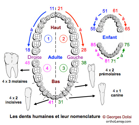 The most commonly used system is the Fédération Dentaire Internationale (FDI) system and this is the one used in Quebec, in Canada and in most European countries.
- Each tooth is represented by a 2-digit number. The upper and lower arch each contains 8 permanent teeth numbered from the midline toward the back from 1 to 8 for the adult dentition.
- Each arch is subdivided in 2 quadrants or semi-arches, a right and left one, indicated by the first digit. The upper right quadrant is #1, and the left one is #2. The lower arch includes quadrants #3 (at the bottom left) and #4 (at the bottom right).
- Teeth in quadrant #1 are numbered starting with “1” followed by the number identifying the tooth (1 to 8). Teeth in quadrant #2 start with “2” and so on for each quadrant.
- Each quadrant contains 8 permanent teeth that are numbered in the following manner:
- Incisors: upper right (11, 12), upper left (21, 22), lower left (31, 32) and lower right (41, 42) (for a total of 8 incisors).
- Canines: upper right (13), upper left (23), lower left (33) and lower right (43) (4 canines).
- Premolars: upper right (14, 15), upper left (24, 25), lower left (34, 35) and lower right (44, 45) (there are 8 premolars).
- Molars: upper right (16, 17), upper left (26, 27), lower left (36, 37) and lower right (46, 47).
- Wisdom teeth (third molars) : upper right (18), upper left (28), lower left (38) and lower right (48) (there are 12 molars).
- (Above illustration reproduced and modified with the author’s permission.)
- To be noted that “Right” and “Left” refer to the patient’s right and left when we look at him in front of us.

Tooth surfaces
Moreover, each tooth has several surfaces:
- Labial: surface on the side of the lips of the anterior teeth (incisors and canines).
- Buccal: surface on the side of the cheeks of the posterior teeth (premolars and molars).
- Lingual: surface on the side of the tongue of every tooth (anterior and posterior).
- Occlusal: masticating surface of the posterior teeth.
- Incisive: sharp edge of the incisors.
- Mésial: interproximal surface (between the adjacent teeth) located nearest to the midline.
- Distal: interproximal surface (between the adjacent teeth) located farthest from the midline.
Universal dental numbering system (United States)
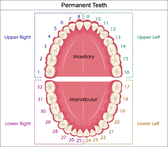
- This numbering system, mainly used in the United States, starts by numbering the teeth at 1 from the upper right part (third molar) and ends at the lower right jaw with tooth #32 (third molar).
- Thus, our tooth #18 is tooth #1 for Americans, tooth #11 is their tooth #9, our tooth #26 is their #15, our #38 is their #19, our #31 is their #26, our #48 is their #36, etc.
- You can imagine the potential confusion if only the tooth numbers were used to communicate between dentists who use different systems. This is why a dental diagram (odontogram) illustrating which teeth we refer to is often included with the description of the dentition in dental work prescriptions (extractions, restorations, surgeries, etc.).

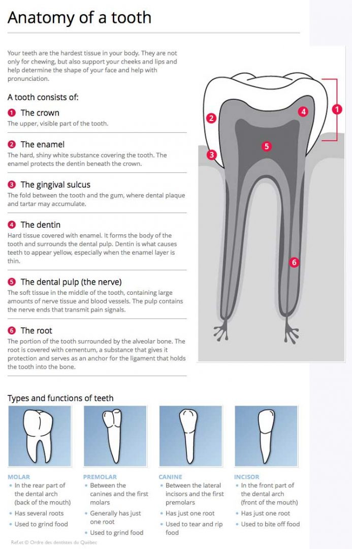
Illustration of the different parts and components of a tooth (lower incisor) and periodontium (alveolar bone and gum tissue).